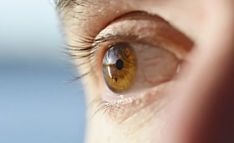
Images of the retina may reveal clues that extend beyond eye diseases. A recent study has found that subtle changes in certain retinal layers are associated with atrial fibrillation, one of the most common heart rhythm disorders. These changes may appear up to four years before the condition is diagnosed clinically.
Atrial fibrillation is a disorder of the electrical activity in the heart’s two upper chambers, known as the atria. It causes an irregular heartbeat and ineffective atrial contractions.


The condition can lead to blood pooling, particularly in the left atrial appendage, increasing the likelihood of blood clots forming. These clots may travel to the brain and cause a stroke.
The US Centers for Disease Control and Prevention reports that atrial fibrillation is associated with an approximately fivefold increase in the risk of ischemic stroke, even after accounting for conventional risk factors.
Global estimates indicate that atrial fibrillation and atrial flutter affected approximately 52.55 million people worldwide in 2021. One of the main challenges with atrial fibrillation is that it can remain undetected because symptoms may be absent or unclear, despite its association with an increased risk of stroke, heart failure, and other complications.
A study published in PLOS Digital Health on September 10 found that 62% of people with atrial fibrillation were unaware they had the condition before receiving a diagnosis. The researchers also reported that changes in a layer of cells at the back of the eye may help identify people who are at risk of developing the disorder up to four years before it is clinically diagnosed.
What Did the Retinal Images Reveal?
Researchers analyzed retinal imaging data from more than 90,000 people across two large British cohorts.
The first group came from the AlzEye database at Moorfields Eye Hospital and included 50,651 people, of whom 5,375 had atrial fibrillation.
The second group was drawn from the UK Biobank. It included 39,013 people who did not have atrial fibrillation at the beginning of the follow-up period. Over an average follow-up of four years, 838 participants were subsequently diagnosed with the condition.
The researchers used optical coherence tomography (OCT), a non-invasive imaging technique that uses light to produce detailed images of the retinal layers, alongside color photographs of the back of the eye.
The findings showed differences in several retinal layers among people with atrial fibrillation. The most consistent result was thinning of the macular ganglion cell–inner plexiform layer (mGCIPL), a neural layer that plays an important role in transmitting visual information from the retina toward the brain.
Notably, people who did not have atrial fibrillation when their eyes were examined but were diagnosed with the condition later had a thinner mGCIPL layer, on average, approximately four years before their clinical diagnosis.
After accounting for factors including age, sex, blood pressure, diabetes, smoking, and alcohol consumption, each standard-deviation decrease in the thickness of this layer was associated with a 27% increase in the subsequent rate of atrial fibrillation.
The findings do not prove that retinal thinning causes atrial fibrillation, nor do they mean that an eye examination can diagnose the heart condition on its own. Instead, they point to a potential way of identifying people who may benefit from additional cardiac assessments.
How Can the Eye Reveal Clues About Heart Health?
Dr. Ehab Saad Osman, professor of ophthalmology and eye surgery at Cairo University’s Faculty of Medicine, explains that the retina is closely connected to the central nervous system. Its tiny blood vessels also provide doctors with a unique opportunity to observe how systemic diseases affect the circulation.
He notes that OCT devices can measure retinal layer thickness with a high degree of precision. This has made the technology an important tool for diagnosing and monitoring eye conditions, including glaucoma and diseases affecting the retina and optic nerve.
The retina consists of multiple interconnected layers. These transfer signals from light-sensitive cells to nerve cells, which process visual information and transmit it to the brain through the optic nerve.
The new findings are significant because consistent changes in the thickness of certain neural layers may reflect early effects of circulatory disturbances or other systemic conditions before the disease becomes clinically apparent.
Dr. Khaled Farouk, professor of critical care medicine at Cairo University’s Kasr Al-Ainy Faculty of Medicine, points out that atrial fibrillation can occur intermittently, with episodes appearing and disappearing. As a result, an electrocardiogram may show a normal rhythm if it is performed when an episode is not occurring.
He adds that some people experience no symptoms, while others may develop palpitations, fatigue, shortness of breath, dizziness, or chest pain. These symptoms are not unique to atrial fibrillation, which can make the condition more difficult to recognize.
A Heart Condition That Can Remain Undetected
The difficulty of identifying atrial fibrillation does not mean that everyone with the condition is unaware of it. However, limited awareness remains a significant challenge.
In a separate survey conducted by the American Heart Association in 2025, involving 770 patients with atrial fibrillation, 62% said they had not known about the condition before being diagnosed. This finding came from an independent awareness study, not from the retinal imaging research.
Early detection is particularly important because treatment may include medications to control heart rate or regulate heart rhythm. Anticoagulants may also be prescribed to patients who need them to reduce the risk of blood clots and stroke.
Can Tiny Blood Clots Leave Traces in the Retina?
The researchers proposed one possible explanation for the relationship between atrial fibrillation and retinal thinning: circulatory disturbances or tiny blockages may reduce blood and oxygen supply to retinal cells, potentially leading to the gradual loss of neural tissue.
However, this explanation remains a hypothesis that requires further investigation. The study did not establish that microscopic blood clots directly caused the retinal changes observed.
Other findings also suggest a possible link between eye health, atrial fibrillation, and its complications. A previous study involving 169 people with atrial fibrillation found that the presence of retinal ischemic perivascular lesions (RIPLs) was associated with a history of stroke. The association remained after adjustment for several known risk factors, with an odds ratio of 2.59.
This result does not mean that these lesions can independently predict who will experience a stroke. The study was cross-sectional and relatively small. Nevertheless, it adds to the evidence suggesting that the retina’s tiny blood vessels may contain information about the state of circulation throughout the body.
Although the findings open a new avenue of research into the potential role of eye imaging in detecting cardiovascular disorders early, further studies are needed before these indicators can become an established screening tool. Researchers must confirm their accuracy, determine how reliably they predict the condition, and establish how they could complement conventional cardiac tests.